
Japan Simulator Trains Surgeons Without Animal Tissue
A new training simulator from Japan lets surgeons practice delicate cancer procedures on realistic artificial tissue, eliminating the need for animal models while keeping patients safer. The device even mimics bleeding and complications to prepare doctors for real emergencies.
Surgeons learning to remove early stomach and colon cancers now have a safer way to practice, thanks to a breakthrough training tool from Japan that feels just like operating on real patients.
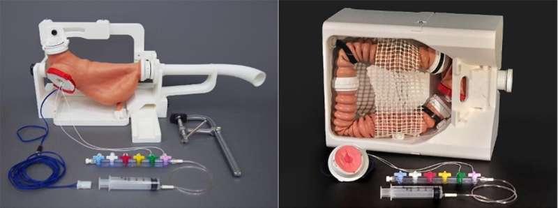
Researchers at Tohoku University partnered with two Japanese companies to create a simulator for endoscopic submucosal dissection, a minimally invasive procedure that removes cancerous tissue through a scope. The technique was invented in Japan but remains difficult to master because one wrong move can cause dangerous bleeding or accidentally pierce the organ wall.
Until now, surgeons typically learned on live animals or practiced directly on patients under supervision. Both approaches have serious drawbacks: animal training raises ethical concerns and hygiene issues, while on-the-job learning puts real patients at risk during a doctor's learning curve.
The new simulator uses specially designed soft materials layered to mimic human tissue. Associate Professor Takeshi Kanno and his team built sheets that recreate the mucosa, submucosa, and muscle layers that surgeons must navigate during the procedure. The materials respond to surgical tools with the same resistance and texture as human tissue.
What makes this simulator truly revolutionary is its ability to teach through mistakes. Artificial blood vessels built into the model start "bleeding" when cut, forcing trainees to practice their emergency response. If a surgeon accidentally damages the muscle layer, a fat layer becomes visible, just like in a real perforation, so doctors can recognize and handle complications before they ever face them in an operating room.
Five expert physicians tested the simulator and confirmed it provides an experience nearly identical to actual surgery. Beginners can now learn core techniques in a risk-free environment, while experienced surgeons can rehearse especially challenging cases before attempting them on patients.
The Ripple Effect
This innovation arrives at a critical time for global healthcare. Endoscopic cancer removal offers patients faster recovery and fewer complications than traditional surgery, but the technique has spread slowly because of training barriers. By standardizing high-quality instruction without animal models or patient risk, this simulator could help doctors worldwide master the procedure.
The device also represents a shift toward more ethical medical education. For decades, medical schools and training programs have sought alternatives to animal testing, and this simulator proves that synthetic materials can now match biological tissue in both feel and function.
Japan's health ministry has already shown interest in the technology for nationwide training programs. The simulator's creators expect hospitals and medical schools around the world to adopt it, potentially training thousands of surgeons who will go on to treat early cancers before they become life-threatening.
The future of surgical training looks like this: doctors fully prepared before their first real incision, patients protected from preventable complications, and medical education that respects both human and animal lives.
More Images


Based on reporting by Medical Xpress
This story was written by BrightWire based on verified news reports.
Spread the positivity! 🌟
Share this good news with someone who needs it


