3-Minute MRI Breakthrough Detects Heart Failure Earlier
Scientists developed a quick, painless MRI scan that measures heart oxygen levels in just three minutes, potentially transforming how doctors catch and treat heart failure early. The technology could help millions of Americans get better care without invasive procedures.
Doctors just got a powerful new tool to spot heart failure before it becomes life-threatening.
Researchers at Cedars-Sinai Medical Center have created a three-minute add-on to standard MRI scans that measures how efficiently your heart uses oxygen. Published this week in Science Translational Medicine, the breakthrough could change care for the 1 in 4 Americans who will develop heart failure in their lifetime.
Until now, checking heart oxygen levels meant inserting catheters into blood vessels or using radioactive tracers with PET scans. Both methods are invasive, expensive, and can't be repeated often to track treatment progress.
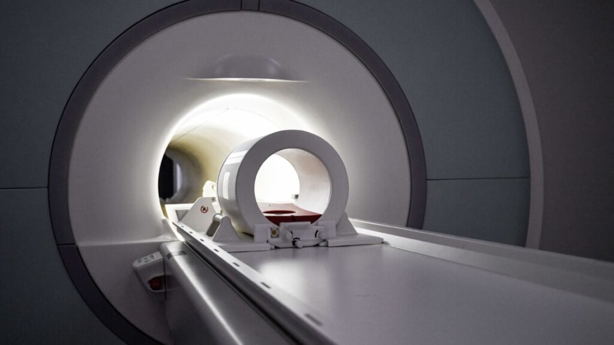
The new approach adds just three minutes to a routine cardiac MRI. No needles, no breath-holding, no radiation exposure.
"A cardiac MRI scan is a routine clinical procedure that people can order," said study co-author Hsin-Jung Yang, an associate professor at Cedars-Sinai. "This is a three-minute add-on, without contrast agent, without breath-holding."
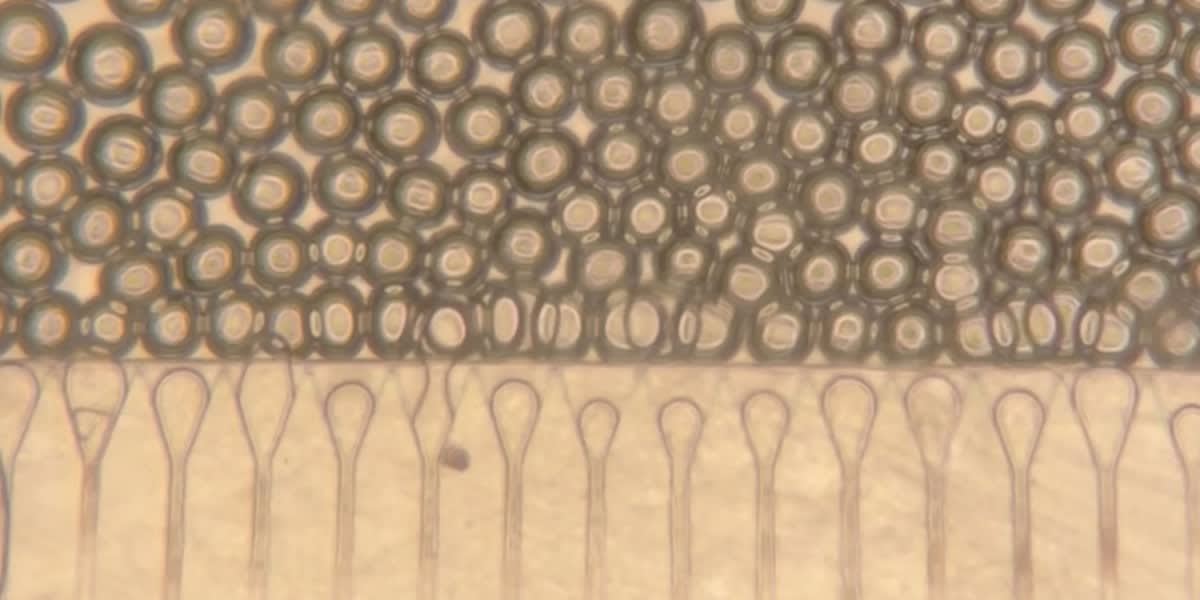
The team first adapted brain imaging technology to capture oxygen signals from beating hearts, a technical challenge that required correcting for constant motion and signal noise. They tested it in pigs, then in 22 heart attack patients, successfully matching results from older invasive methods.
The timing couldn't be better. A growing number of people have heart failure with preserved ejection fraction, a condition where the heart pumps normally but still doesn't deliver enough blood. Doctors are now treating it with obesity drugs like GLP-1s and SGLT2 inhibitors, which show promising results for metabolic problems in the heart.
This new scan could help doctors see exactly how those treatments are working. Because it's noninvasive and uses no contrast agents, doctors can repeat it safely to monitor patient progress over time.
Why This Inspires
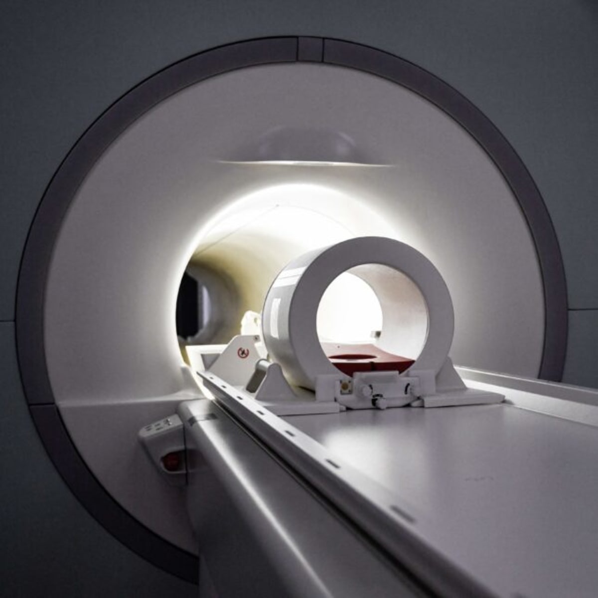
Heart failure affects millions, but catching it early has always been complicated and uncomfortable. This breakthrough transforms a dreaded diagnostic journey into something as simple as lying in an MRI tube for a few extra minutes.
Every major hospital already has MRI machines, which means this technology could reach patients quickly once approved. Lower-risk populations who couldn't undergo catheter procedures can now get tested. Doctors can check treatment response regularly without putting patients through repeated invasive tests.
The research team is already collaborating with colleagues on complementary approaches, including metabolite imaging that tracks other signs of heart health. Together, these advances paint a picture of a future where heart failure gets caught early, treated precisely, and monitored easily.
One quick scan could mean the difference between catching heart problems early and waiting until symptoms become severe.
More Images


Based on reporting by STAT News
This story was written by BrightWire based on verified news reports.
Spread the positivity!
Share this good news with someone who needs it