AI Tool Predicts Brain Age and Cancer Survival from MRIs
Researchers at Mass General Brigham created an AI tool that can analyze brain scans to predict everything from dementia risk to cancer survival, even with limited training data. The breakthrough could help doctors provide faster, more personalized care to patients worldwide.
A new artificial intelligence tool is transforming how doctors read brain scans, opening doors to faster diagnoses and better patient care.
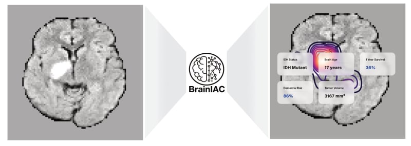
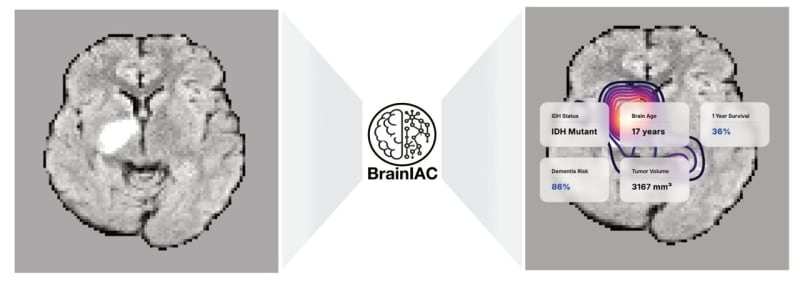
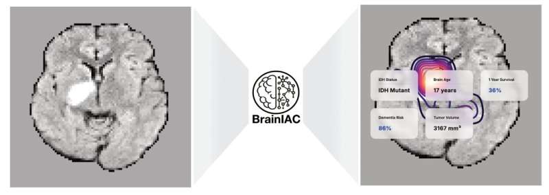
Researchers at Mass General Brigham developed BrainIAC, an AI model that can analyze brain MRI images to perform multiple medical tasks at once. The tool can estimate brain age, predict dementia risk, detect brain tumor mutations, and forecast cancer survival rates.
What makes BrainIAC special is its ability to learn from unlabeled medical images. Most AI tools need thousands of carefully labeled scans to work properly, but this one teaches itself to recognize patterns first, then applies that knowledge to specific medical tasks.
The research team tested BrainIAC on nearly 49,000 brain scans across seven different medical challenges. It successfully handled both simple tasks like classifying scan types and complex ones like identifying specific tumor mutations.
BrainIAC outperformed three other specialized AI models designed for single tasks. It proved especially valuable when training data was scarce, a common problem in medical research where annotated datasets can be hard to find.
The Ripple Effect
This breakthrough could accelerate medical discoveries that previously took years. When AI can analyze diverse brain scans without needing extensive labeled data, smaller hospitals and research centers gain access to powerful diagnostic tools they couldn't build themselves.
Dr. Benjamin Kann, who led the research at Mass General Brigham's Artificial Intelligence in Medicine Program, believes integrating BrainIAC into standard imaging protocols could help clinicians personalize treatment plans faster. Patients could receive more accurate diagnoses earlier in their care journey.
The tool works across different types of brain images, whether they come from neurology departments or cancer centers. This flexibility matters because medical images vary widely between institutions, often making it hard for AI systems to adapt.
The findings, published in Nature Neuroscience, represent a shift toward more versatile medical AI. Instead of building separate tools for each medical question, researchers are creating foundation models that adapt to whatever doctors need.
The team plans to test BrainIAC on additional imaging methods and larger patient populations. As the technology improves, it could become a standard part of how radiologists and neurologists evaluate brain health.
Better diagnostic tools mean patients get answers faster, and doctors can focus their expertise where it matters most.
More Images


Based on reporting by Medical Xpress
This story was written by BrightWire based on verified news reports.
Spread the positivity!
Share this good news with someone who needs it