Lab-Grown Cells Restore Vision in Mice with Retinal Disease
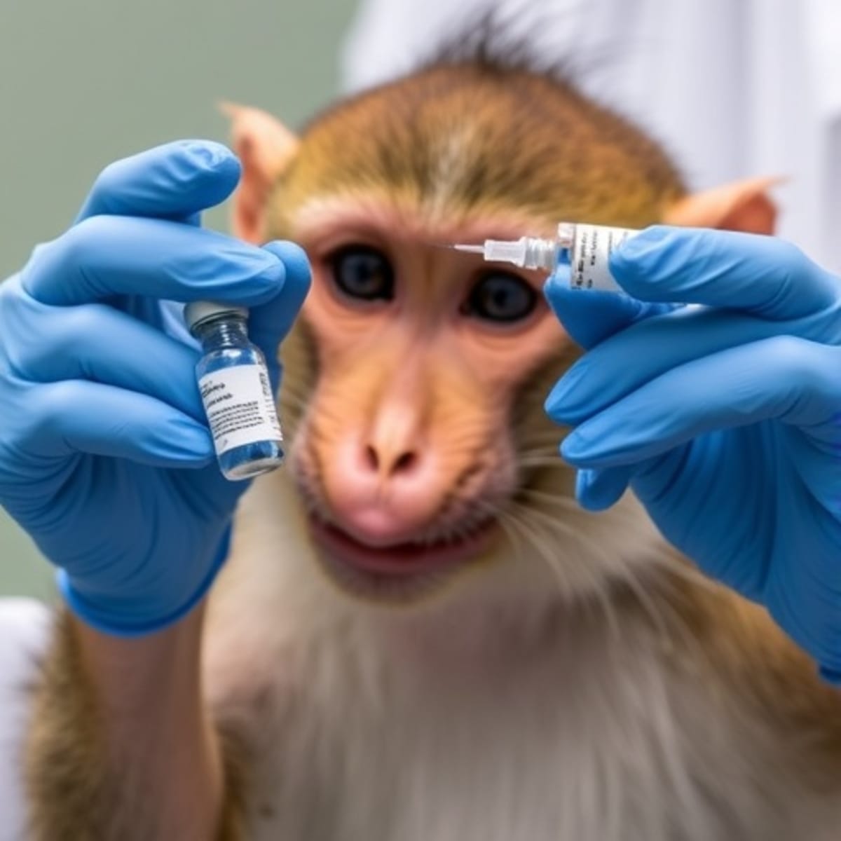
Scientists at Duke University grew specialized retinal blood vessel cells from stem cells and successfully used them to restore vision in mice with damaged retinas. The breakthrough could lead to new treatments for millions suffering from diabetic retinopathy and other causes of blindness.
Scientists just took a major step toward curing blindness by growing the exact cells needed to repair damaged retinas and proving they work in living animals.
Researchers at Duke University in North Carolina created specialized blood vessel cells that keep retinas healthy using stem cell technology. When they injected these lab-grown cells into mice with retinal disease, something remarkable happened: the cells integrated perfectly into damaged tissue, regenerated blood vessels, and restored retinal function.
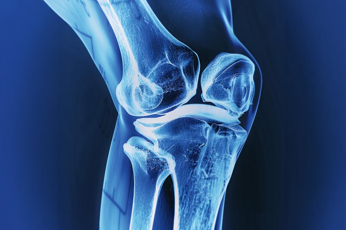
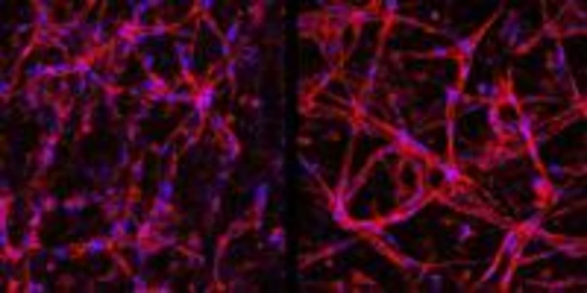
The team focused on retinal endothelial cells, which form the inner layer of blood vessels in the eye. These cells create a critical barrier that controls what enters and exits the retina, including oxygen, nutrients, and medications. When this barrier breaks down, it causes vision loss in millions of people.
Lead researcher Sharon Gerecht explains why treating retinal diseases has been so challenging. The retina connects directly to the brain and has a protective blood barrier that keeps it healthy but also blocks most treatments from getting through.
The breakthrough solves a major problem in eye disease research. Previously, these specialized cells could only be collected from real patients, making them expensive and limited in supply. The Duke team figured out how to grow unlimited quantities from induced pluripotent stem cells, which are mature adult cells reprogrammed to become blank slates that can develop into almost any cell type.
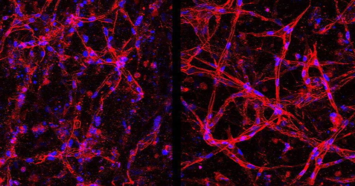
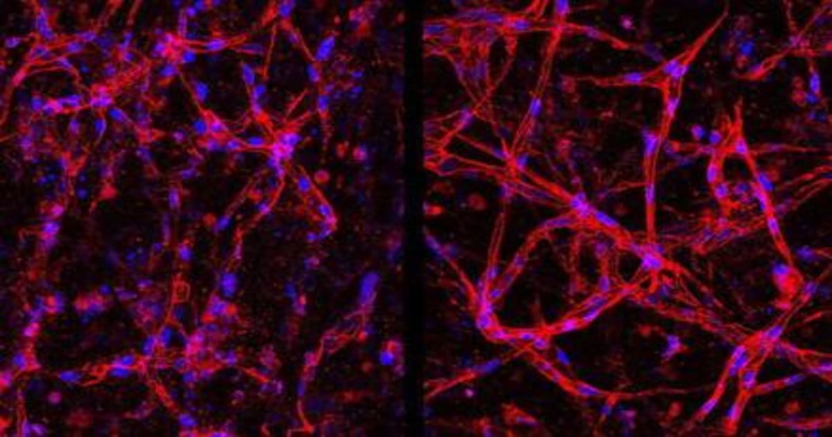
Graduate student Parker Esswein and his colleagues started with commercial stem cells and guided them through a careful process using specialized growth factors. First they coaxed them into common blood vessel cells, then into the specific type found only in retinas.
The researchers put their creation through rigorous tests. They grew the cells into networks matching real retinal tissue, then exposed them to low oxygen and high glucose conditions that mimic diabetic retinopathy. The tissue barrier broke down exactly like it does in patients, proving the lab model works for studying disease.
Why This Inspires
The real magic happened when scientists tested the cells as an actual therapy. Mice with weak, poorly structured retinal blood vessels received injections of the lab-grown cells before vision loss occurred. The cells didn't just survive; they integrated seamlessly into existing tissue and helped build strong, healthy blood vessels with proper protective barriers.
This success opens doors that were previously locked. Diabetic retinopathy alone causes most vision loss in working-age Americans, affecting millions who desperately need better treatment options. Now researchers have both an unlimited supply of the exact cells needed for therapies and a lab model to test new approaches safely before trying them in people.
The technique makes these precious cells easier and cheaper to obtain, potentially expanding access to treatments for patients worldwide. While human trials remain in the future, the foundation is set for transformative therapies that could preserve or restore sight for countless people facing blindness.
Every medical breakthrough starts with a single successful experiment, and this one just proved that science can now grow the building blocks of vision itself.
More Images


Based on reporting by Google News - Health Breakthrough
This story was written by BrightWire based on verified news reports.
Spread the positivity!
Share this good news with someone who needs it