MIT Creates At-Home Breast Cancer Scanner Anyone Can Use
MIT engineers built a portable ultrasound device that creates 3D breast images and requires zero medical training to operate. The smartphone-sized scanner could catch aggressive tumors that develop between annual mammograms.
A new handheld ultrasound device could help catch breast cancers that slip through the cracks of yearly screenings, and you won't need a medical degree to use it.
MIT researchers have developed a portable breast cancer scanner that's barely bigger than a smartphone and simple enough for anyone to operate at home. The device creates detailed 3D images of breast tissue by scanning just two or three spots, guided by an intuitive computer interface that shows users exactly where to place it.
The technology targets a critical gap in cancer care. Between 20 and 30 percent of breast cancers are "interval cancers" that develop between annual mammograms. These tumors tend to be more aggressive and harder to treat because they're caught later.
Associate professor Canan Dagdeviren started this project after losing her aunt to an interval breast cancer in 2015. She wanted something that worked better for women with dense breast tissue and could be used far more often than traditional mammography.
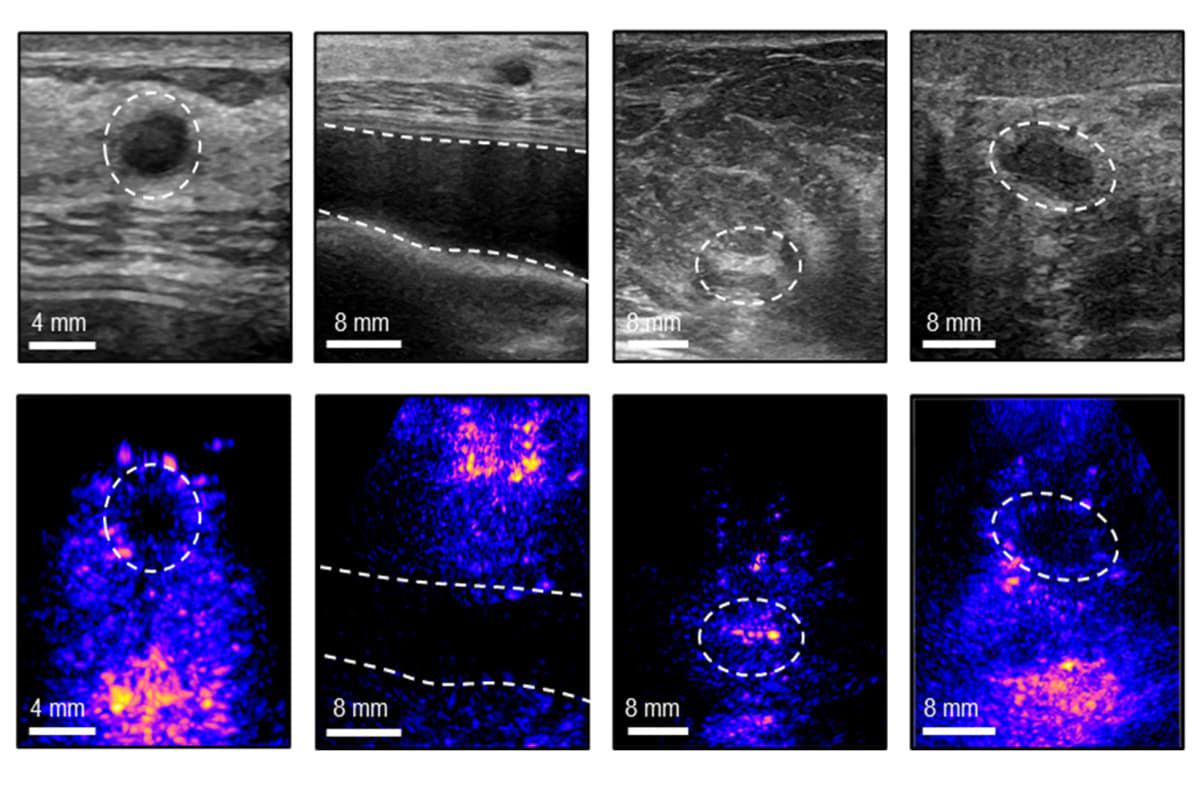
The team's latest breakthrough, published in Nature Communications, includes several key improvements. They added a special "backing layer" to the ultrasound transducer that focuses sound waves more precisely, creating sharper images with less noise. They also designed an algorithm that adjusts for how sound travels differently through various tissues like skin and fat.
Former MIT postdoc Md Osman Goni Nayeem explains the backing layer produces "more accurate and sharper images, with a wider operating range of frequencies." The result is clearer pictures that make it easier to spot potential tumors, cysts, and microcalcifications.
The user interface sets this device apart from traditional ultrasound equipment. The computer guides users to position the probe in exactly the same location each time, which is essential for tracking changes in tissue over weeks or months. No specialized training required.
The Ripple Effect
This technology could transform breast cancer monitoring for millions of women. Those at high risk could check themselves weekly or monthly instead of waiting a full year between screenings. Cancer survivors could track their tissue long-term after treatment, either at their doctor's office or in the comfort of home.
The portable design also means better access in underserved areas where ultrasound equipment and trained technicians are scarce. Rural clinics and community health centers could offer regular breast imaging without major equipment investments.
By putting powerful diagnostic tools directly in patients' hands, MIT's device represents a shift toward proactive, frequent monitoring instead of periodic checkups that might miss fast-growing cancers.
Early detection saves lives, and now that early detection could happen in your living room.
Based on reporting by MIT News
This story was written by BrightWire based on verified news reports.
Spread the positivity!
Share this good news with someone who needs it