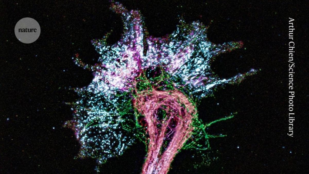
Scientists Inflate Cells 1,000x to Map Proteins on $10K Scope
Researchers have made biological samples one billion times bigger using the same material found in diapers, letting ordinary microscopes see details once reserved for million-dollar equipment. The breakthrough could bring cutting-edge protein research to labs worldwide.
Scientists just figured out how to inflate cells to the size of mouse brains, and it's making the invisible visible for everyone.
Using a super-absorbent polymer similar to what makes diapers work, researchers have expanded biological samples 1,000 times larger in each direction. That's a billion-fold increase in volume, enough to blow up a dime-sized tissue sample to the size of an Olympic swimming pool.
The real magic? This massive expansion lets regular light microscopes see individual amino acids, the building blocks of proteins that were previously only visible with equipment costing millions of dollars.
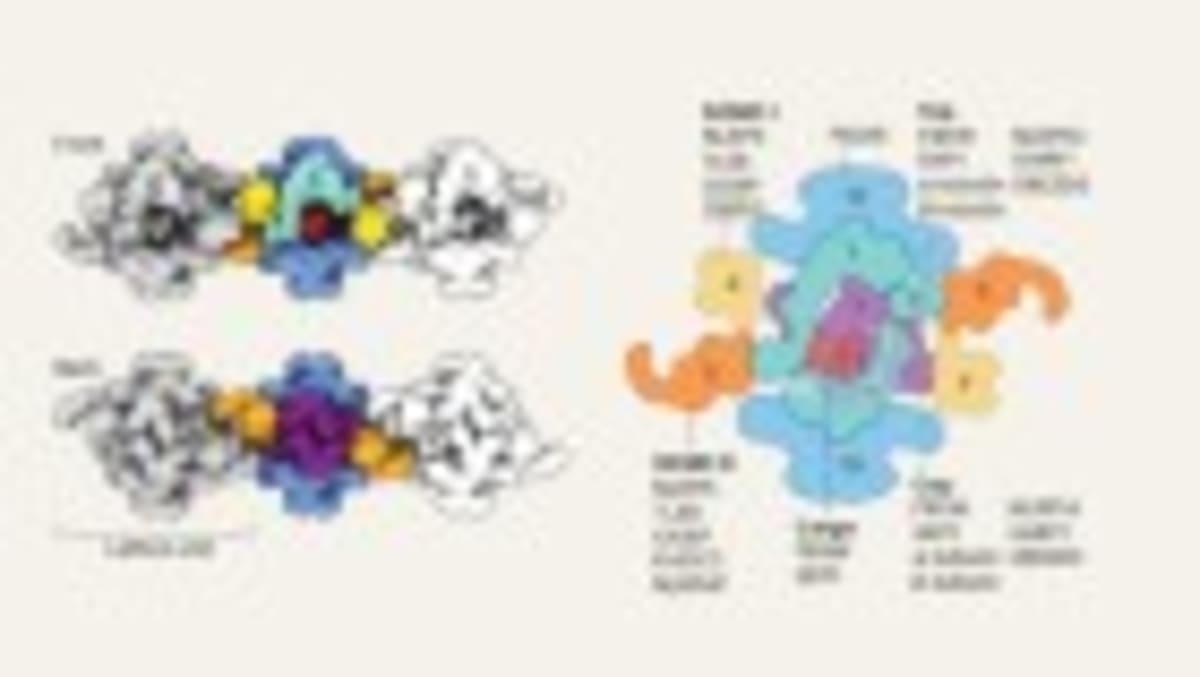
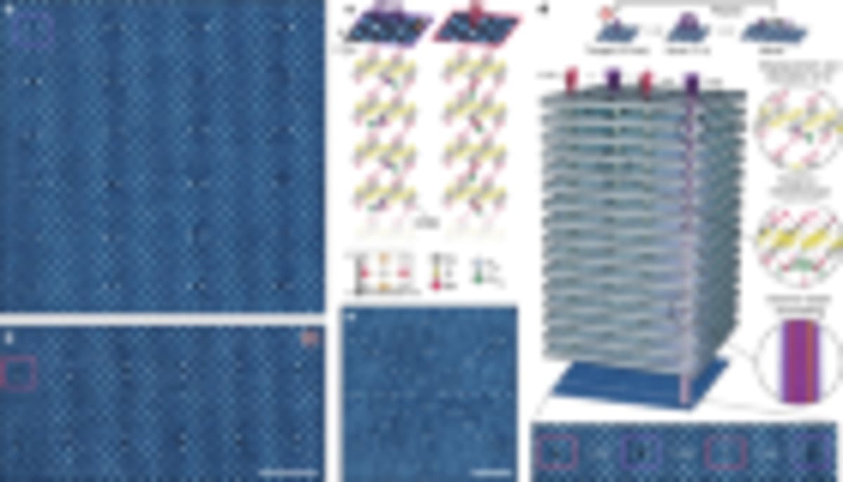
The team from MIT and the University Medical Center Göttingen tested their method on several proteins, including green fluorescent protein, a lab staple. They reconstructed its structure from thousands of snapshots, achieving resolution of about 1.2 nanometers using a standard fluorescent microscope.
The technique builds on "expansion microscopy," invented in 2015 by MIT neuroengineer Edward Boyden. Previous methods could only expand samples about 20 times, but the new approach inflates them multiple times using an improved hydrogel recipe.
Here's how it works: proteins are bonded to the gel, then carefully broken apart with enzymes or heat. This stretches them without destroying their 3D shape, letting scientists map where each amino acid sits.
The researchers have already mapped the structure of mCLING, a nine-amino-acid peptide, and a nanobody (a miniature antibody). Their measurements matched structures predicted by computer simulations.
The Ripple Effect
The democratization of structural biology is now within reach. For decades, mapping protein structures required cryogenic electron microscopy or X-ray crystallography, techniques that demand specialized facilities and deep pockets.
Now labs with basic equipment can explore protein structures at near-atomic resolution. Co-author Silvio Rizzoli notes the team can already determine protein shapes inside living cells at around 10 angstroms, approaching the detail level of cryo-EM.
The potential extends far beyond cost savings. Smaller research institutions, universities in developing countries, and teaching labs could soon access structural biology that was previously out of reach.
The team continues refining their technique, with unpublished work already pushing resolution below 10 angstroms for purified proteins. Co-author Ali Shaib says they're "trying to make cryo-EM like structures from affordable optical microscopes."
This breakthrough opens doors to understanding diseases, developing better drugs, and training the next generation of scientists, all with equipment that fits on a standard lab bench.
More Images

Based on reporting by Nature News
This story was written by BrightWire based on verified news reports.
Spread the positivity!
Share this good news with someone who needs it