Scientists Map Clitoris Nerves in 3D for First Time
Dutch researchers used advanced X-ray imaging to create the first complete 3D map of the clitoris's nerve system, revealing surprising complexity that could improve surgical outcomes. The breakthrough closes a knowledge gap that has existed for decades in medical science.
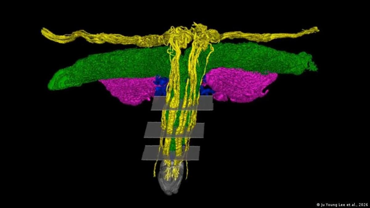
For the first time ever, scientists have mapped the complete nerve system of the clitoris in stunning 3D detail, revealing a complexity that surprises even medical experts.
A research team at Amsterdam University Medical Center used synchrotron radiation, an ultra-high-resolution form of X-ray imaging, to trace the full path of the clitoris's main sensory nerve. What they found challenges previous assumptions and fills a gap that has persisted in medical textbooks for generations.
The images show that inside the clitoral glans, several large nerve trunks branch out in a tree-like pattern toward the surface, some measuring up to 0.7 millimeters in diameter. Contrary to earlier beliefs, these nerves don't simply taper off but form intricate networks that extend beyond the glans into surrounding tissue.
"For the first time, the full trajectory of the terminal nerve branches of the clitoris has been mapped in three dimensions," said Georga Longhurst, division lead of Anatomy and Physiology at the University of London. Previous studies had shown these nerves before, but never with this level of clarity.
The research highlights a longstanding imbalance in medical science. While the penis has been thoroughly documented in textbooks for decades, the clitoris has received far less attention despite the two organs sharing the same embryological origin and similar functions.
Neuroscientist Ju Young Lee, who led the study, recalls asking colleagues at a major conference whether anyone was studying nerves in gynecological organs. The response stayed with her: "Oh, I'd never thought about that." She decided to change that.
Why This Inspires
Since the study was released, surgeons have already reached out to say the findings are helping their daily work. The detailed nerve maps will help doctors operating in the vulva region avoid accidental nerve damage during procedures.
The practical applications span multiple types of surgery: childbirth procedures, gender-affirming surgeries, and reconstructive surgeries following genital mutilation. Each of these procedures requires precise knowledge of nerve pathways to preserve sensation and function.
The research is part of the Human Organ Atlas Hub, an international project systematically mapping the human body using advanced imaging technology. Think of it as creating a Google Earth for human anatomy, ensuring every organ gets the detailed attention it deserves.
Australian urologist Helen O'Connell pioneered similar work in the late 1990s, using MRI to demonstrate that the clitoris measures 8 to 12 centimeters in total length when internal structures are included. Lee's team has now taken that understanding to an unprecedented level of detail.
What makes this particularly meaningful is its simplicity: scientists applied existing technology to an organ that had simply been overlooked, proving that sometimes progress comes not from inventing new tools but from deciding what deserves our attention.
Based on reporting by DW News
This story was written by BrightWire based on verified news reports.
Spread the positivity!
Share this good news with someone who needs it