Singapore Team Shrinks X-Ray Tech Using Graphite Flakes
Scientists just figured out how to create powerful medical X-rays using simple graphite flakes on a table-sized machine. This breakthrough could replace building-sized equipment and make advanced cell imaging accessible to thousands more researchers.
Imagine if the X-ray technology that requires a building-sized machine could fit on a desk. Researchers at Nanyang Technological University in Singapore just made that leap possible using one of the simplest materials on Earth: graphite.
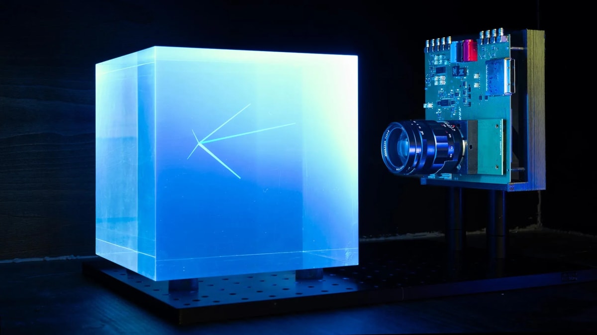
The team discovered that thin flakes of graphite, no thicker than a human hair, can produce what scientists call "water-window" X-rays. These special X-rays let doctors and researchers see living cells in crystal-clear detail without staining or damaging them first.
Here's why this matters for medicine. Current machines that produce these X-rays come in two frustrating flavors: small tabletop versions that only work at fixed settings, or synchrotrons that cost millions of dollars and take up more space than a house. Most hospitals and research labs can't access either option effectively.
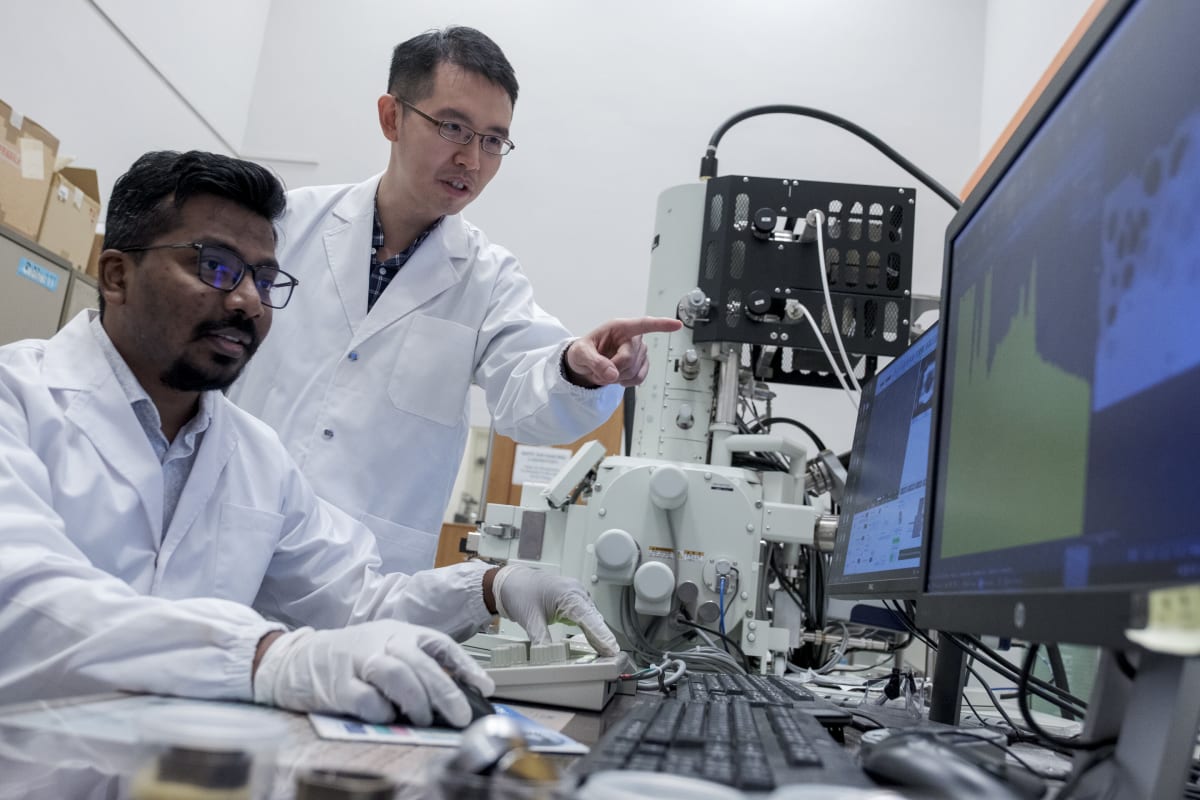
Associate Professor Wong Liang Jie and his team solved both problems at once. Their setup fits on a table and can adjust to different energy levels simply by changing how an electron beam hits the graphite or tilting the material at different angles.
The research, published in Nature Photonics, shows the team didn't just stumble onto this discovery. They developed precise mathematical frameworks to predict exactly how electrons would scatter off crystalline materials, then confirmed their predictions through experiments.
The Ripple Effect
This breakthrough could democratize an entire field of biological imaging. Thousands of research labs currently locked out of synchrotron access could soon afford their own flexible X-ray systems. Medical centers in developing countries could image cells and tissues with technology that was previously out of reach.
The graphite flakes work at thicknesses between 10 and 170 nanometers, materials that are relatively inexpensive and easy to produce. The team's framework for predicting X-ray production also gives other scientists a roadmap to optimize the technology further.
Beyond research labs, this could speed up drug development and disease diagnosis. When more scientists can see cells at high resolution without complex preparation, they can run more experiments, test more treatments, and make discoveries faster.
Making cutting-edge medical technology smaller, cheaper, and more accessible turns an exclusive tool into an everyday resource for saving lives.
More Images


Based on reporting by Phys.org
This story was written by BrightWire based on verified news reports.
Spread the positivity!
Share this good news with someone who needs it