ASU Scientists Find Weak Spot in Dangerous Tick Disease
Arizona State University researchers just cracked a mystery that could lead to new treatments for tularemia, a deadly tick-borne disease so potent that just 10 bacterial cells can cause infection. For the first time, scientists isolated and studied the proteins that help this dangerous pathogen survive inside human cells.
Scientists just found a promising weakness in one of nature's most efficient killers.
Researchers at Arizona State University have achieved something that has eluded scientists for years. They successfully isolated and studied a group of proteins that allow tularemia bacteria to infect human cells and evade our immune defenses.
Tularemia is a rare but serious disease spread by ticks, contaminated water, and even through the air. What makes it terrifying is its efficiency: fewer than 10 bacterial cells can trigger a full infection. The disease causes fever, swollen lymph nodes, and sometimes pneumonia, and without quick treatment with antibiotics, it can be fatal.
Public health agencies classify tularemia as a high priority pathogen because it spreads so easily. It has even been studied as a potential bioweapon in the past.
The breakthrough centers on proteins called the CapBCA complex, which sit inside the bacterium's inner membrane. Scientists have known for years that these proteins are essential for infection. When researchers disabled them in animal studies, the bacteria became much weaker.
But studying these proteins has been nearly impossible because they live embedded in cell membranes. No one had figured out how to produce them, isolate them, or keep them stable in a lab until now.
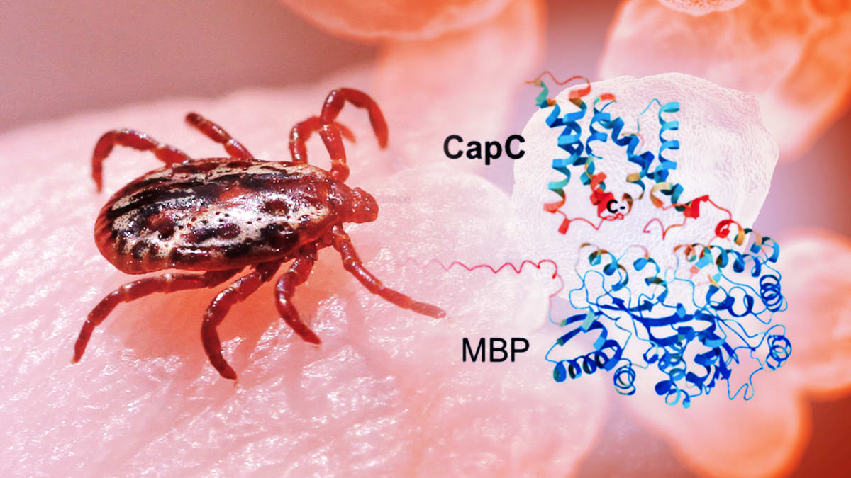
The ASU team, led by Petra Fromme at the Biodesign Center for Applied Structural Discovery, found a clever solution. They inserted the relevant genes into common E. coli bacteria, adding a molecular tag that guided the proteins into cell membranes and made them easier to track. Then they used a gentle detergent to extract the proteins without destroying them.
First author Eranjalee Ranaweera and her colleagues discovered that the proteins are mostly spiral shaped and stick together in small groups rather than working alone. Using imaging and biochemical techniques, they captured the first glimpse of how these protein assemblies form and function.
Why This Inspires
This discovery represents years of patient problem solving paying off. The team overcame technical barriers that had stopped researchers worldwide, opening a door that was firmly locked.
Now that scientists can produce and study these proteins, they can work toward understanding their exact structure and function. That knowledge is the foundation needed to design drugs that target these proteins specifically, potentially stopping the infection before it takes hold.
With antibiotic resistance becoming a growing concern even for tularemia, having new treatment options matters more than ever. This research gives doctors and patients hope for better weapons against a dangerous disease.
The work appears in the journal Biochimica et Biophysica Acta Biomembranes. Fromme says the team is excited to continue revealing how these proteins work together, believing this insight will eventually lead to new therapeutic strategies.
Sometimes the biggest victories come from solving problems others thought were impossible.
Based on reporting by Google News - New Treatment
This story was written by BrightWire based on verified news reports.
Spread the positivity!
Share this good news with someone who needs it