Dr. Lydia Wilson Unveils 4D Scan for Lung Cancer
A breakthrough 4D imaging technique gives cancer surgeons a real-time roadmap of how lungs breathe. This advance helps protect healthy tissue and could unlock life-saving surgery for thousands of vulnerable patients.
A breakthrough in lung imaging is giving cancer patients a brighter, safer path to curative surgery. For individuals facing early-stage lung cancer, this new visual roadmap ensures that vital breathing tissue stays protected when they need it most.
Surgery offers the strongest chance of a cure for early lung cancer, but only if the patient has enough healthy lung capacity to recover safely. Traditional tests often assume lung function is spread evenly across the chest, which can leave doctors guessing in complex cases.
Now, researchers at Thomas Jefferson University have introduced a smarter solution using four-dimensional computed tomography ventilation, or 4DCT-ventilation. Lead researcher Dr. Lydia Wilson and her team adapted this radiation technology to create dynamic, moving maps of patient breathing.
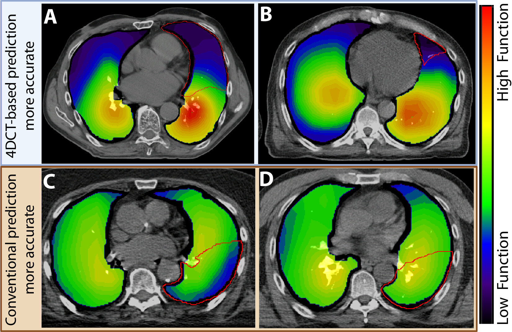
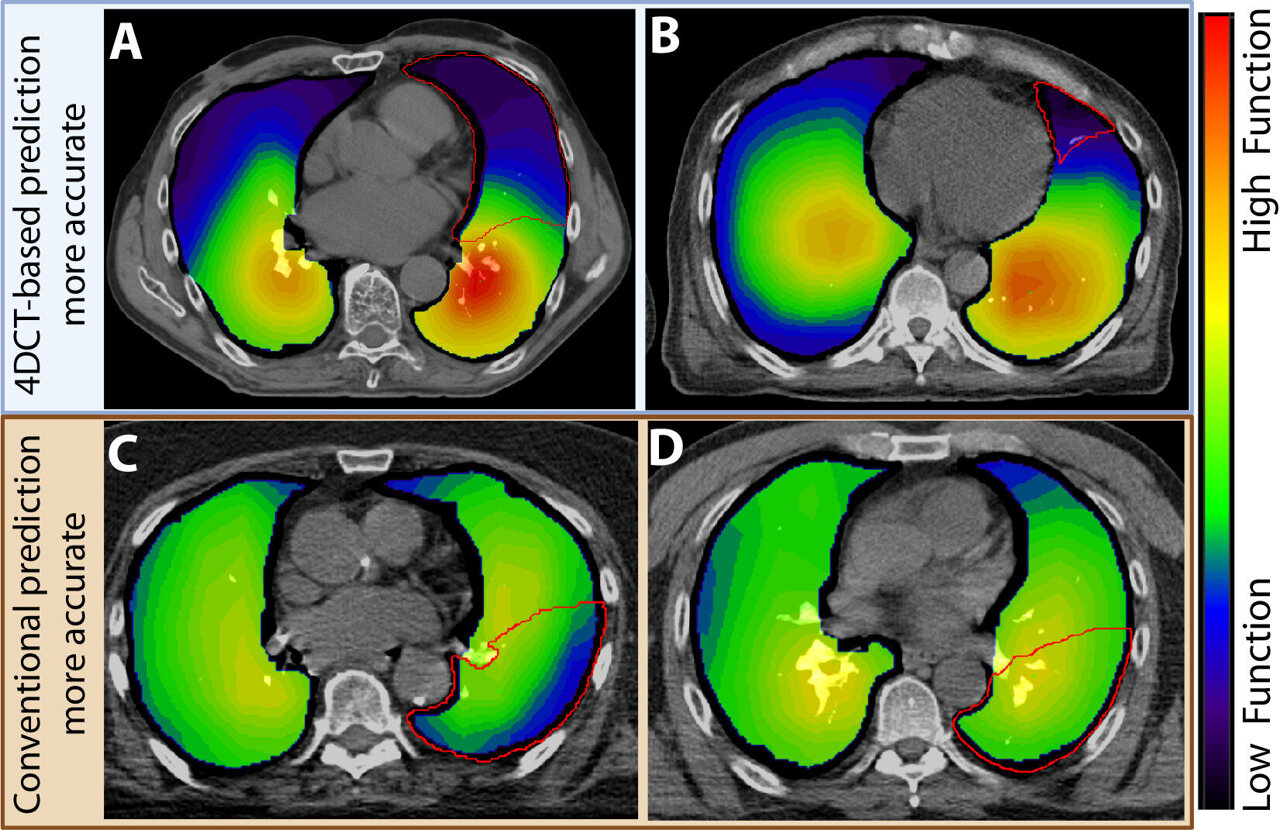
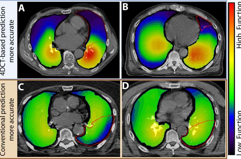
During a standard 4DCT scan, patients simply breathe in and out while the camera captures their real-time respiratory motion. Advanced software transforms that video into a color-coded map that reveals exactly which sections of the lung are thriving and which are struggling.
This precision is a game-changer for people with pre-existing conditions like emphysema or chronic obstructive pulmonary disease. In these patients, lung strength is rarely uniform, making surgical planning far more delicate.
Removing the wrong section could accidentally take away a patient's only functioning tissue. By contrast, seeing the precise working zones helps surgeons remove tumors while safeguarding the healthy areas that keep patients breathing easily.
The team evaluated data from 30 early-stage lung cancer patients to test their technique against standard methods. When researchers reviewed the outcomes months later, the 4D scans proved remarkably accurate, especially for patients with uneven lung performance.
Unlike traditional alternatives, this gentle imaging method requires no contrast dyes and demands no grueling physical stress from the patient. It delivers clear answers rapidly while keeping patient comfort front and center.
Dr. Wilson is already planning the next phase of research to evaluate patients previously considered borderline or ineligible for surgery. Her team believes this tool can expand surgical access to people who were once told their lungs were too weak for an operation.
The Bright Side
This breakthrough turns a standard scan into a powerful navigation system that replaces guesswork with crystal-clear vision. As precision medicine advances, thousands of high-risk patients can look forward to safer procedures, quicker recoveries, and a renewed lease on life.
Every breath tells a story, and this new window into our lungs is helping more cancer survivors breathe easy.
More Images
Based on reporting by Medical Xpress
Reset Your Focus With Today’s Daily Affirmations & Mood Check
Take a 60-second mindful pause with curated daily affirmations and our private positivity mood tracker.
Spread the positivity!
Share this good news with someone who needs it