MIT Creates "X-Ray Vision" Ultrasound Using VR Headset
MIT researchers turned ultrasound imaging into a 3D experience using virtual reality headsets, letting doctors see inside the body like they have X-ray vision. The breakthrough could make medical training faster and diagnoses more accurate.
Imagine a doctor putting on a VR headset and instantly seeing a 3D image of what's happening inside your body, floating right where it should be in real space.
MIT researchers just made that possible with a new ultrasound system that transforms the confusing black-and-white images we're used to into crystal-clear 3D representations. Using augmented reality, medical professionals can now see through skin and tissue as if they have superpowers.
The old way of doing ultrasounds required serious mental gymnastics. Technicians had to look at flat, 2D images on a screen and mentally piece them together to understand what they were really seeing inside the body. It's a difficult skill that takes years to master, and even experts can miss things.
Jason Hou, one of the lead researchers and an MIT graduate student, calls it a "mental tomography bottleneck." That cognitive burden can lead to mistakes and missed diagnoses.
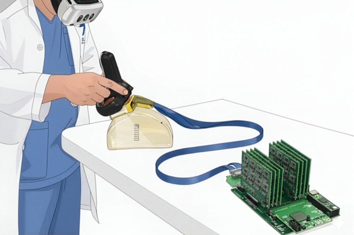
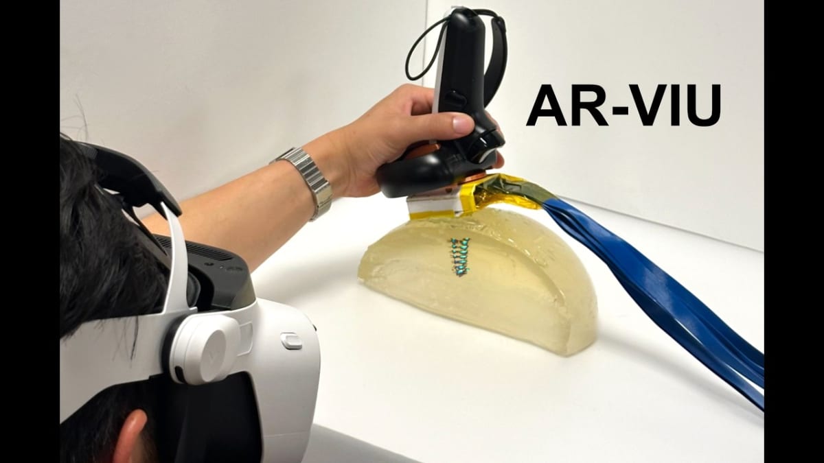
The new system, called AR-VIU, uses a compact ultrasound probe about the size of a deck of cards. It captures 3D images in real time and streams them into a gaming engine called Unreal Engine, which converts the data into a viewable 3D model. When users wear an AR headset, they see the internal structures superimposed exactly where they exist in the body.
The magic happens when users move their heads or walk around. The 3D image shifts with their perspective, just like looking at a real object from different angles. It's intuitive in a way traditional ultrasound never could be.
The research team tested their technology with 18 people, split evenly between ultrasound experts and complete beginners. They asked participants to identify objects in gelatin using four different methods: traditional 2D ultrasound, 3D ultrasound on a regular screen, 2D augmented reality, and the full 3D AR system.
The results were striking. Everyone performed better with the AR-VIU system, but the biggest winners were the beginners. They could identify objects almost as accurately as the experts, suggesting the technology could dramatically shorten training time.
The Ripple Effect
This breakthrough could reshape medical training and patient care in powerful ways. New ultrasound technicians could learn in months instead of years, addressing healthcare worker shortages. Doctors performing delicate procedures like biopsies could place needles with more confidence and precision.
Associate professor Canan Dagdeviren, who led the study, sees even deeper benefits. "On the clinical side, it could be less time-consuming, more accurate, and also give health care providers more peace of mind," she says. "They wouldn't have to wonder if they missed anything."
The system costs less to build than traditional 3D ultrasound because it uses fewer components and requires less power. That means more hospitals and clinics could afford the technology, bringing better care to more patients.
The study appeared in Nature Communications Engineering, marking another win for combining cutting-edge gaming technology with medicine to solve real problems and save real lives.
More Images
Based on reporting by MIT News
This story was written by BrightWire based on verified news reports.
Spread the positivity!
Share this good news with someone who needs it