MIT's AI Makes Surgeries Safer in Seconds
Researchers created an AI system that helps surgeons navigate minimally invasive procedures with sub-millimeter precision in just five minutes. The breakthrough could make life-saving surgeries accessible to millions more Americans.
Imagine a surgeon threading a tiny catheter through your arteries to save your life, guided only by grainy X-ray images that look like shadowy puzzles. Now MIT researchers have created an AI system that turns those puzzles into crystal-clear roadmaps in seconds.
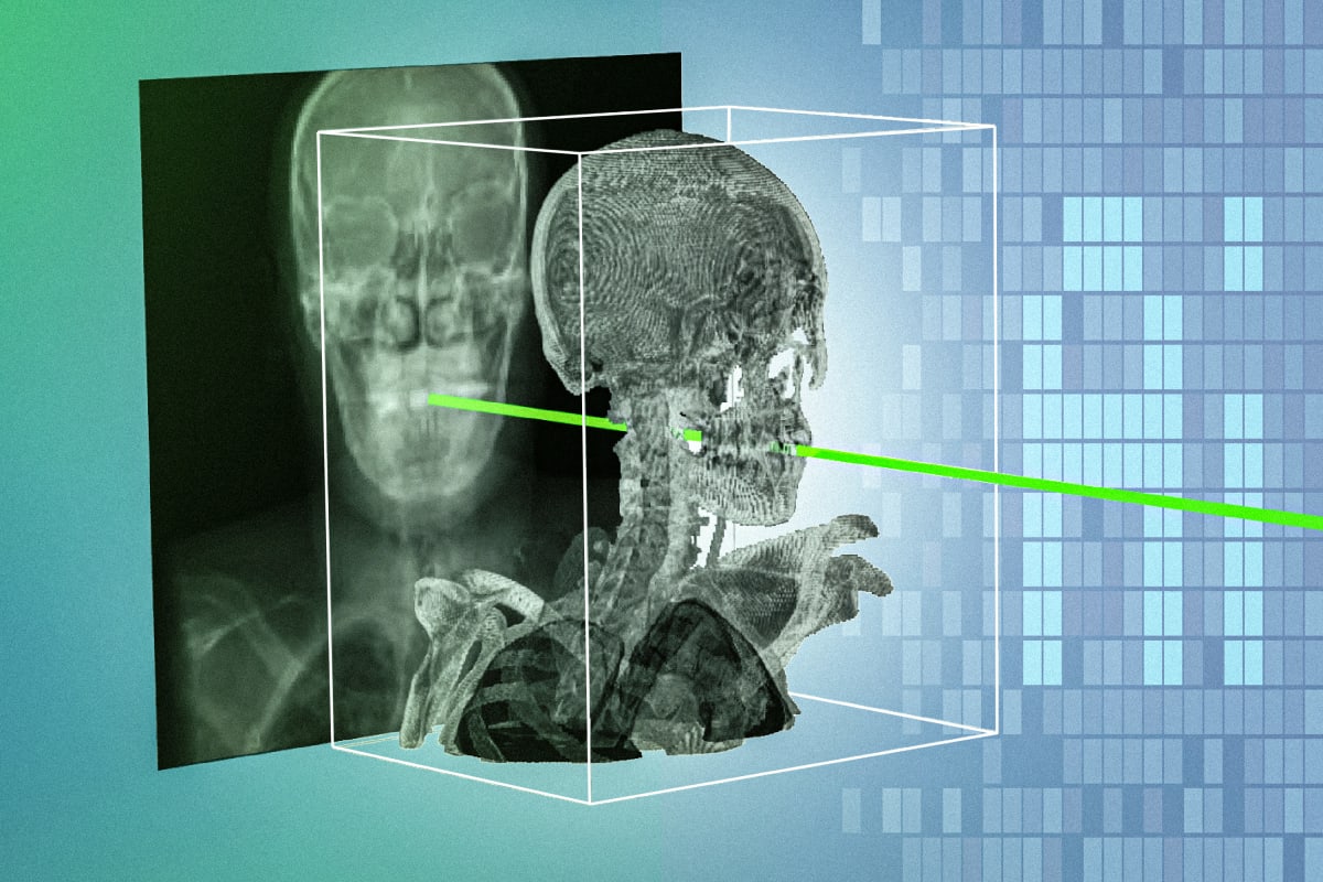
The new technique, called xvr, matches real-time surgical X-rays with a patient's 3D medical scans automatically and with incredible accuracy. It adapts to each patient in about five minutes and works ten times better than existing AI methods.
Right now, surgeons performing minimally invasive procedures face a tricky challenge. They use X-rays to see inside the body during surgery, but X-rays are flat images that make it hard to know exactly where surgical tools are located. Manual methods to solve this problem are slow and require clinicians to guess positions by clicking landmarks on a screen.
The MIT team, led by postdoc Vivek Gopalakrishnan, built an AI model that generates thousands of synthetic X-rays from different angles using a patient's preoperative CT or MRI scan. The system then matches these with real X-rays captured during surgery, pinpointing the exact location and orientation of surgical instruments.
The technology works across different patients, body parts, and medical procedures. It performed with sub-millimeter precision in testing, meaning surgeons can guide their tools with exceptional accuracy through tiny incisions.
The Ripple Effect
This breakthrough matters most for people living far from specialized medical centers. A majority of Americans live more than an hour away from facilities that perform procedures like emergency stroke interventions, where every minute counts.
"An hour in stroke time is incredibly substantial," Gopalakrishnan explains. Making these highly specialized procedures easier means they become accessible to much broader parts of the population.
The team published their findings in Nature and tested the system on real surgical procedures. Collaborators include clinicians from Harvard Medical School, Massachusetts General Hospital, Boston Children's Hospital, and other leading institutions.
The researchers designed xvr specifically to handle the incredible diversity of human anatomy. Previous AI tools struggled because they couldn't adapt robustly to different patients, making them impractical in actual operating rooms.
By combining 2D X-rays with 3D medical imaging information seamlessly, xvr gives surgeons superpowers they've never had before. It transforms procedures that once required decades of training to visualize properly into operations guided by precise, real-time mapping.
The system processes about 1,000 synthetic X-ray images each second, making it fast enough for real surgical environments where speed matters as much as accuracy.
This innovation could reshape how we think about access to advanced medical care, bringing specialized life-saving procedures within reach of hospitals and patients who previously couldn't access them.
Based on reporting by MIT News
This story was written by BrightWire based on verified news reports.
Spread the positivity!
Share this good news with someone who needs it