MRI Breakthrough Detects Brain Damage at Microscopic Level
Scientists have transformed standard MRI machines into powerful microscopes that can spot brain damage at the cellular level, years before symptoms appear. This medical breakthrough could revolutionize how doctors diagnose and treat conditions like traumatic brain injury, Alzheimer's, and multiple sclerosis.
Imagine detecting brain damage before you ever feel a symptom, giving doctors years of precious time to intervene and potentially save your cognitive health.
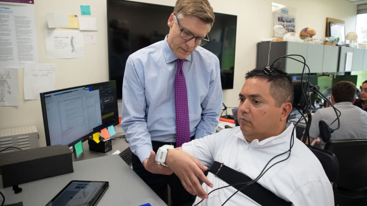
Researchers at the University of Eastern Finland and New York University just made that possible. They've discovered how to turn ordinary MRI machines into precision tools that can detect microscopic changes in brain tissue, something that was previously impossible without invasive procedures.
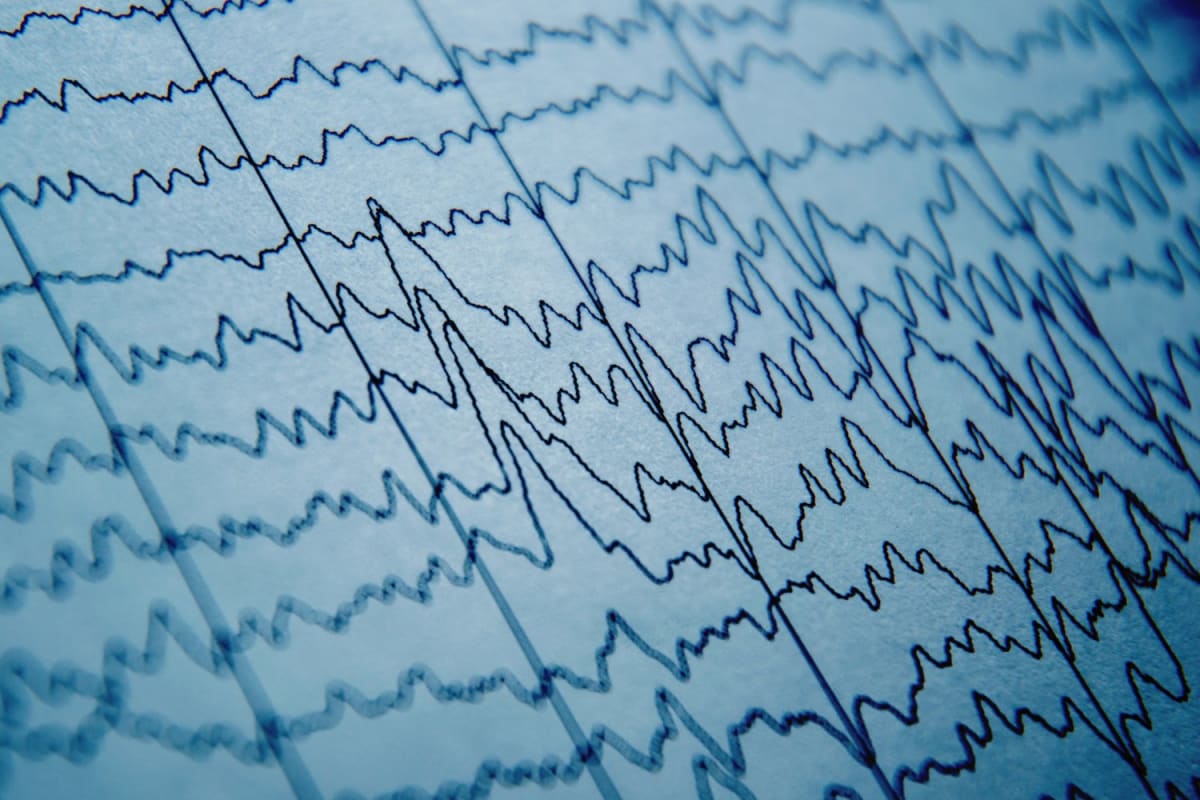
The breakthrough focuses on white matter, the brain's internal wiring system made up of axons that transmit electrical signals between neurons. When these structures get damaged, it's often the first sign of neurological disorders, but traditional imaging couldn't see these tiny changes until they became severe.
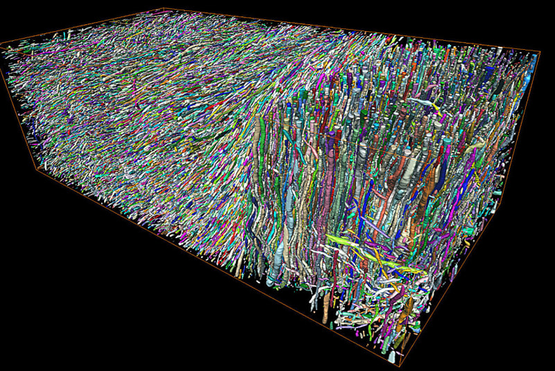
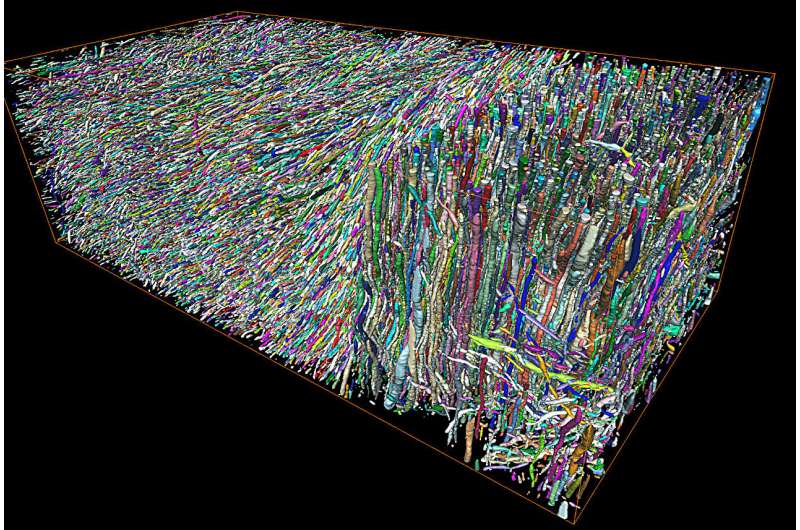
Dr. Ali Abdollahzadeh and his team solved this puzzle by combining two cutting-edge technologies. They used electron microscopy to create the world's largest 3D map of brain white matter, reconstructing hundreds of thousands of axons with nanometer precision. Then they figured out the mathematical relationship between what MRI scanners see and what's actually happening at the cellular level.
The result is remarkable. Standard diffusion MRI, which measures how water moves through tissue, can now quantify specific structural changes in axons. The team validated their approach using rat models of traumatic brain injury, detecting microscopic damage months after injury occurred.
Why This Inspires
This isn't just about better diagnostics. It's about hope for millions living with or at risk for neurological conditions.
Early detection means early intervention. Doctors could potentially slow or stop disease progression in conditions like multiple sclerosis, Alzheimer's, and Parkinson's before irreversible damage occurs. Athletes could be monitored for cumulative brain trauma with unprecedented precision. Treatments could be tested and refined based on actual cellular-level changes, not just whether symptoms improve.
The technology doesn't require new MRI machines or invasive procedures. It works with existing equipment, meaning hospitals worldwide could adopt it relatively quickly. Teams are already translating the approach to human brain imaging at Kuopio University Hospital and NYU's new state-of-the-art scanner.
Professor Els Fieremans captures the magnitude: "The combination of animal model research and clinical MRI creates a translational path from nanometer-scale tissue to human neuroimaging, allowing us to test these biomarkers in patients for the first time."
The path from microscopic tissue damage to a doctor's diagnosis just got infinitely shorter, and that changes everything for brain health.
More Images

Based on reporting by Medical Xpress
This story was written by BrightWire based on verified news reports.
Spread the positivity!
Share this good news with someone who needs it