New 3D Color Scan Sees Inside Body Without Radiation
Scientists at Caltech and USC created a medical imaging system that produces vivid 3D color images of tissue and blood vessels in under a minute, without radiation or dyes. The breakthrough could transform how doctors detect cancer, monitor nerve damage, and study the brain.
Imagine getting a detailed medical scan that shows your doctor both the structure of your organs and how blood flows through them, all in living color and without a single dose of radiation.
That future just arrived. Researchers at Caltech and USC have developed a new imaging technique that combines ultrasound with light-based technology to create stunning 3D color images of the human body. The system, called RUS-PAT, has already been tested on human volunteers and is moving toward clinical use.
The breakthrough addresses a longstanding problem in medical imaging. Standard ultrasound is fast and affordable but only shows flat, grayscale images of tissue shape. Other advanced scans like CT and MRI require contrast dyes, expose patients to radiation, or take too long for frequent monitoring.
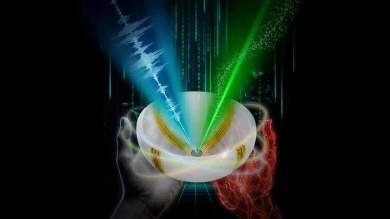
Professor Lihong Wang, who leads the project, found an elegant solution by merging two technologies in a completely new way. His team uses laser light to trigger tiny sound waves inside tissue, which reveals how blood vessels function. At the same time, ultrasound waves map out the physical structure. Arc-shaped detectors rotate around the scanning area, capturing both signals at once.
The entire scan takes less than one minute. It works on tissue up to 4 centimeters deep, and doctors can reach even deeper areas using endoscopic tools.
Why This Inspires
This technology could change lives in remarkably practical ways. Breast cancer patients could get scans that not only locate tumors but also show their biological activity, helping doctors choose the best treatment. People with diabetes could monitor both nerve damage and blood flow in a single appointment, catching problems before they become serious.
Dr. Charles Liu, a neurosurgeon at USC who collaborated on the project, sees enormous potential for brain research. Scientists could watch blood flow patterns while studying brain anatomy, opening new windows into how our minds work.
The system costs less to build than current advanced imaging equipment because it uses just a handful of detectors instead of hundreds. That affordability could help more hospitals and clinics offer cutting-edge diagnostic care.
Wang's innovation came from a simple question: instead of using expensive arrays of ultrasound transducers, what if one device could do both jobs? That moment of curiosity led to technology that addresses the limitations of nearly every major imaging method in use today.
The human trials have already begun, with volunteers and patients providing the first real-world proof that the system works safely and effectively.
Medical imaging has spent decades getting better at seeing inside us, but this approach does something more: it shows us in motion, in color, and in ways that could help doctors catch diseases earlier and treat them smarter.
Based on reporting by Science Daily
This story was written by BrightWire based on verified news reports.
Spread the positivity!
Share this good news with someone who needs it