New Cell Imaging Method Could Transform Disease Detection
Scientists just made it easier and cheaper to peek inside our cells and understand how our bodies respond to diet and disease. This breakthrough technique could revolutionize how doctors diagnose and treat illnesses.
Your body is constantly adapting to everything you eat, every stress you face, and every challenge you encounter. Now, scientists have found an affordable new way to watch that adaptation happen in real time inside individual cells.
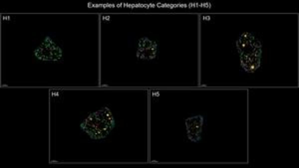
Researchers at HHMI's Janelia Research Campus developed a technique called Spatial Organellomics that examines the tiny structures inside cells called organelles. Think of organelles as a cell's internal organs. Just like your heart and lungs respond to exercise, organelles change shape and behavior based on what's happening around them.
The breakthrough is that this method doesn't require expensive specialized equipment or destroy tissue samples. Scientists can now create an "organelle signature" for every cell in a tissue, mapping how cells change their internal machinery to handle different conditions.
The team tested their approach on liver cells and discovered something unexpected. Instead of being organized in neat zones as scientists previously thought, different types of liver cells were mixed throughout the organ. This intermingling might actually help the liver adapt better to challenges like toxins or dietary changes.
When researchers fed mice different diets, one low in nutrients and another high in fat and carbs, the liver cells completely reorganized themselves. Each diet created unique cell types, but with less variety than before. This finding suggests that extreme diets might reduce the liver's ability to fight off future threats like infections or cancer.
The team then trained AI models to predict cell states and even identify what diet an animal was eating just by looking at organelle signatures. The accuracy rate exceeded 95 percent. The method can also track fatty liver disease progression, pointing toward future diagnostic applications.
Why This Inspires
This research represents more than just scientific curiosity. By understanding how our cells respond to environmental stresses at such a detailed level, doctors could eventually catch diseases earlier and design more effective treatments tailored to each person's unique cellular responses.
The team is already expanding beyond liver research. They're collaborating with other labs to examine cells in the gut and pancreas, opening doors to understanding digestive diseases and diabetes in entirely new ways.
What makes this particularly exciting is the accessibility. Because the technique doesn't require expensive equipment, more research labs around the world can use it to study how tissues respond to everything from medications to environmental toxins.
Our bodies are constantly working to keep us healthy, making millions of tiny adjustments we never notice. Now scientists can finally see that remarkable adaptation in action, one cell at a time.
Based on reporting by Google News - Researchers Find
This story was written by BrightWire based on verified news reports.
Spread the positivity!
Share this good news with someone who needs it