Scientists See Living Cancer Cells Like Never Before
Researchers just made cancer studies easier with new dyes that let scientists watch living cells in action without damaging them. The breakthrough could speed up discoveries in how cancer spreads and responds to treatment.
Scientists can now watch cancer cells move and change in real time, thanks to microscope dyes that blink naturally like fireflies.
A team led by Oregon Health & Science University researcher Catherine Galbraith just published a breakthrough that makes cutting-edge microscopy accessible to more cancer labs. The new fluorescent dyes, developed with Howard Hughes Medical Institute's Luke Lavis, solve a problem that's frustrated researchers for years.
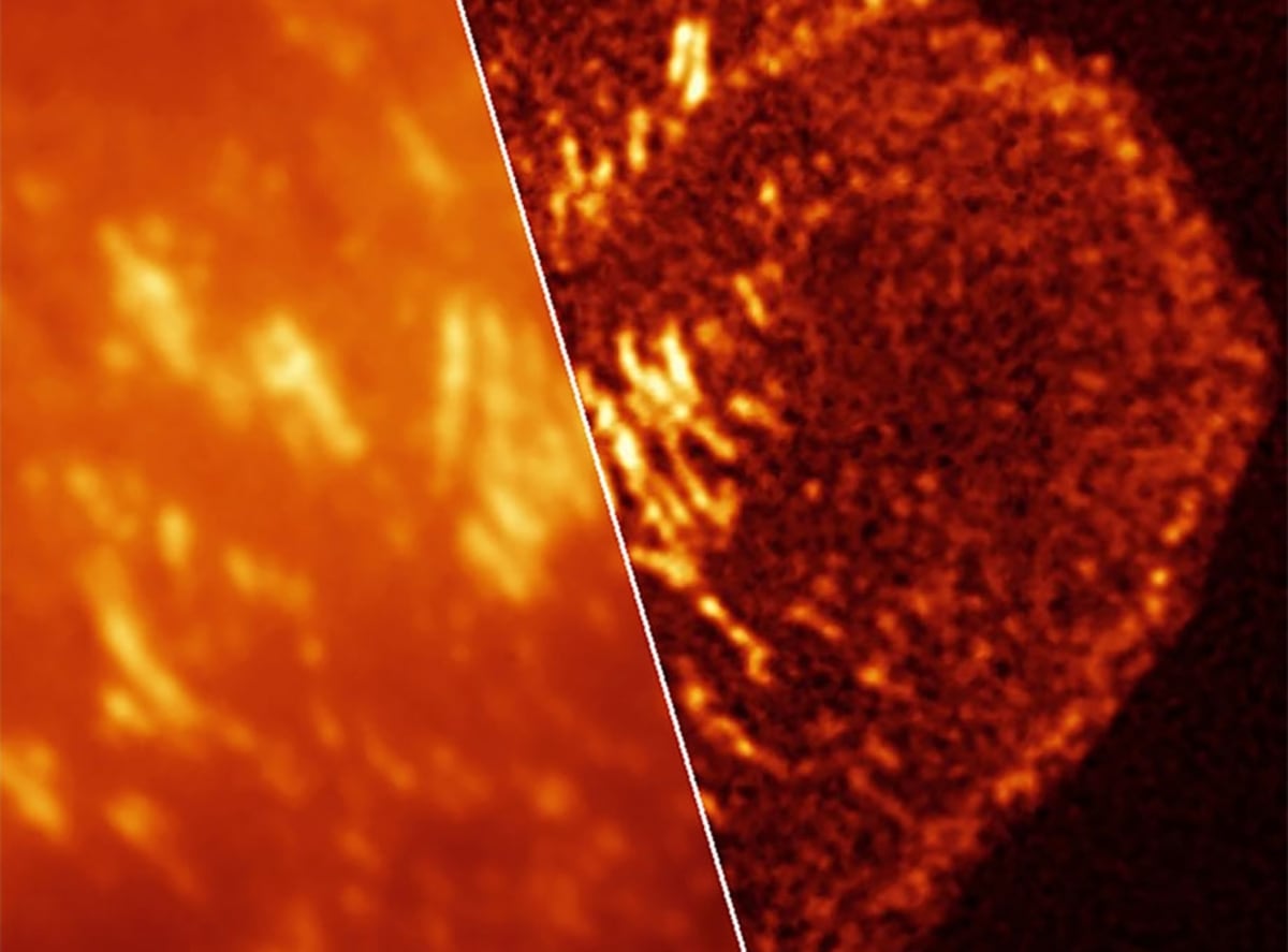
Super-resolution microscopy lets scientists see tiny structures inside cells that regular microscopes miss entirely. But using this powerful technique on living cells has been notoriously difficult, requiring intense light or harsh chemicals that often kill the cells scientists are trying to study.
The newly developed dyes change that game completely. They flicker on and off naturally, without needing special equipment or damaging the cells they illuminate.
"Luke's dyes remove a major barrier that has kept live super-resolution imaging out of reach for many biology labs," Galbraith said. Her team figured out which dyes work best in different situations, creating a practical guide for researchers.
The timing couldn't be better for cancer research. Tumor cells constantly shift how they grow, move, and resist treatment, and understanding those changes requires watching them happen live.
The new tools let researchers observe critical processes tied to cancer: how DNA packages and unpacks, how genes turn on and off, how cancer cells migrate through tissue, and how molecules organize inside cells. These are the small-scale decisions that determine whether cancer spreads or responds to therapy.
The Ripple Effect
The research, published in Nature Methods, represents Galbraith's second paper in a Nature journal in just two weeks. But the real impact goes beyond academic prestige.
Cancer labs that couldn't afford specialized equipment or didn't have expertise in complex imaging techniques can now study living cells at unprecedented detail using standard microscopes. The dyes work with existing technology most labs already own.
Lavis's Janelia Fluor dyes are already used in microscopy labs worldwide, and researchers expect these blinking versions to spread just as quickly. Galbraith created a framework showing exactly which dye performs best in living cells, fixed cells, or the acidic environments found inside tumors.
The tools open doors to questions researchers couldn't answer before. Watching a cancer cell grip and invade tissue in real time reveals details that still images of dead cells simply can't capture.
"New tools open up biology," Galbraith said, and this one arrives when cancer researchers need better ways to understand their constantly changing opponents.
More Images

Based on reporting by Google News - Researchers Find
This story was written by BrightWire based on verified news reports.
Spread the positivity!
Share this good news with someone who needs it