Indian Scientists Map Human Brainstem in 3D at Cellular Level
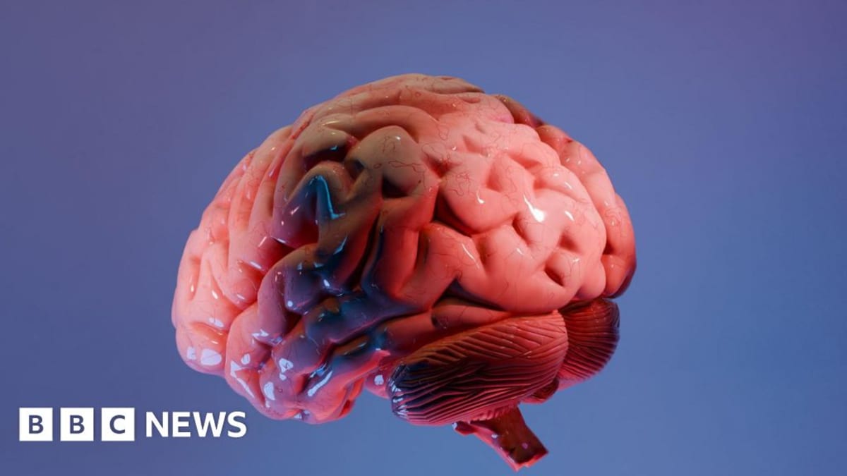
Scientists in India have created the world's most detailed 3D atlas of the human brainstem, bridging the gap between brain scans and individual cells. The free online tool could transform how doctors understand and treat Alzheimer's, Parkinson's, and stroke.
For over a century, doctors studying brain diseases like Alzheimer's have examined only 15 to 20 tissue slices from an organ containing 86 billion neurons. Now, Indian scientists have created a breakthrough map that lets researchers travel from whole brain scans down to individual nerve cells without losing their place.
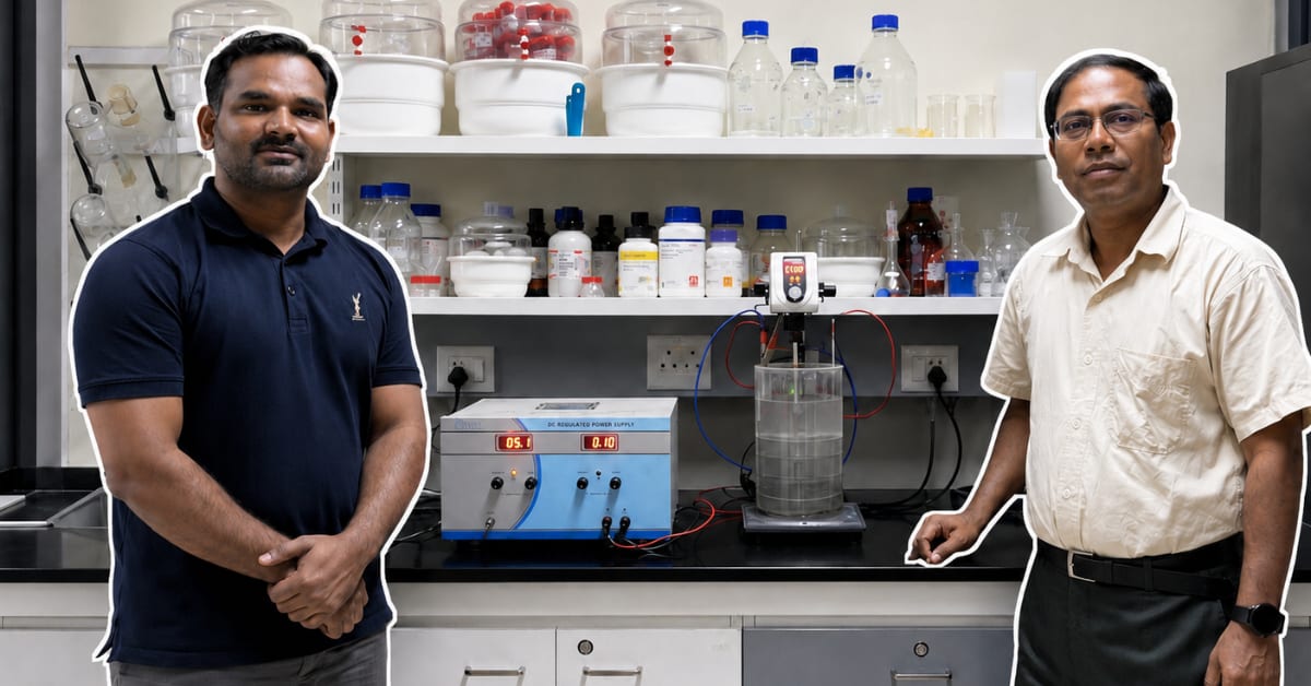
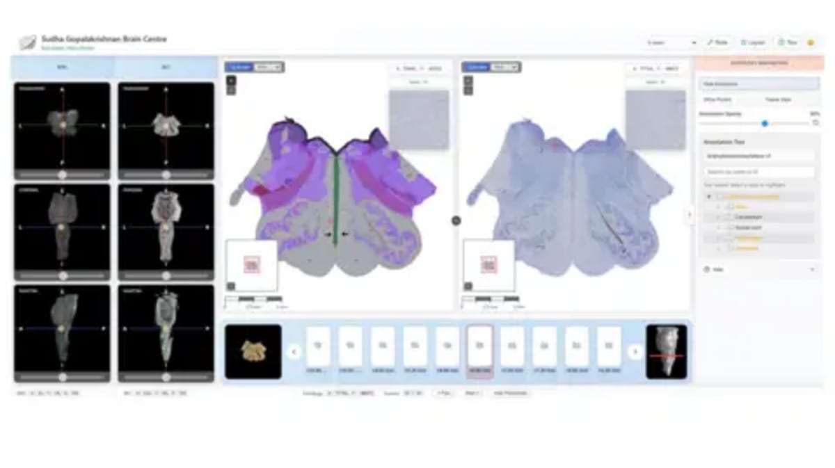
The team at the Sudha Gopalakrishnan Brain Centre in Chennai built the world's most detailed 3D atlas of the human brainstem. Called Anchor, it combines more than 500 tissue sections from brains at different life stages into one seamless digital map that anyone can explore online for free.
The brainstem may be tiny, but it keeps us alive. This sliver of tissue controls breathing, heartbeat, sleep, and movement. Damage to even small clusters of cells can prove catastrophic, yet its densely packed structure has frustrated mapping efforts for decades.
What makes Anchor special is how it connects two worlds that rarely meet. Medical imaging shows the whole brain but misses cellular detail. Microscopes reveal individual cells but only in isolated slices. Anchor bridges that gap, identifying more than 200 clusters of brain cells and nerve pathways with stunning clarity.
The project took 18 months and about 20 scientists working together. They used high-resolution microscope images rather than expensive molecular techniques, making detailed brain mapping affordable for the first time at this scale.
Dr. Rebecca Folkerth, who has examined thousands of brains over three decades at Harvard Medical School and NYU, calls it a dream come true. "What the Indian centre has created is essentially what I dreamed of early in my career," she told the BBC.
Why This Inspires
Scientists worldwide can now access this atlas free online, opening doors to discoveries that seemed impossible just years ago. By comparing healthy brainstem maps with diseased tissue, researchers may finally understand why Parkinson's develops, how strokes damage brain tissue that might still be saved, and what triggers sudden infant death syndrome.
The atlas could help neurosurgeons navigate one of the brain's most delicate regions with greater confidence. Early findings have already uncovered new features that could help doctors preserve injured brain tissue during strokes, potentially improving patient outcomes.
Brain scientist Partha Mitra from Cold Spring Harbor Laboratory says detailed atlases like this could have a "transformative impact" on neurological disease research. The tool could even help explain how infections like Covid-19 trigger long-term brain damage.
International neuroscientist Shubha Tole describes the project as putting India "at the international table" with an "unprecedented integration" of engineering, neuroscience, and medicine. The achievement reflects how modern neuroscience increasingly depends on computation and engineering as much as biology.
Twenty scientists working together have mapped what was once considered unmappable, and they're sharing it freely with the world.
More Images

Based on reporting by Google News - Health
This story was written by BrightWire based on verified news reports.
Spread the positivity!
Share this good news with someone who needs it