Laser Breakthrough Makes Protein Imaging Sharper Than Ever
After 15 years of skepticism, scientists finally cracked a technology that lets microscopes see tiny proteins in stunning detail. The laser phase plate could unlock mysteries about how proteins work inside living cells.
Scientists just proved the doubters wrong with a breakthrough that transforms how we see the building blocks of life.
For 15 years, structural biologists argued over whether a laser-based imaging tool could ever actually work. Many experts said it was impossible. This month, two research teams proved it wasn't just possible but revolutionary, publishing results that show the laser phase plate (LPP) dramatically improves how we image proteins.
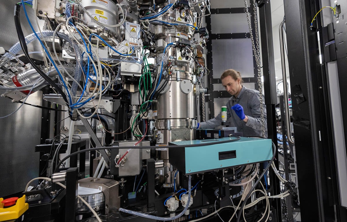
The technology addresses a tricky problem in cryo-electron microscopy, where scientists freeze proteins in ice to photograph them. Proteins don't naturally absorb electrons, making them nearly invisible under microscopes. They scatter electrons instead, which researchers have struggled to detect clearly.
Biophysicist Robert Glaeser and physicist Holger Müller started exploring a wild idea in 2010 at UC Berkeley. They wanted to focus an intense laser onto the electron beam to boost image contrast. When Müller presented early prototypes to cryo-EM specialists, he remembers the reaction clearly: "The sentiment was, 'This would be so cool, but this will never work.'"
The challenges were enormous. The team needed to create the brightest continuous laser focus anywhere on Earth, using lasers that didn't even exist when they started. Their solution involved precisely machined mirrors so smooth their surface roughness measures less than the diameter of a hydrogen atom.
These mirrors reflect an incoming laser back and forth thousands of times, amplifying it to the necessary intensity. Made from special ceramic glass, they resist damage from the intense beam while maintaining long-term stability.
The Ripple Effect
The breakthrough extends cryo-EM to smaller proteins that were previously too difficult to image clearly. It also simplifies tomography experiments that show how proteins behave inside actual cells, not just in isolation.
Biohub in Redwood City, California, funded the project's development after seeing its potential. David Agard, founding scientific director of imaging at Biohub, calls it "the first exciting new thing to happen in cryo-EM hardware beyond things that have been around for decades now."
The technology opens doors to understanding diseases at the molecular level and designing better treatments. Being able to see smaller proteins with greater clarity means scientists can study biological processes that were previously invisible, from how viruses infect cells to how our bodies build and repair tissue.
After years of skepticism and technical obstacles that seemed insurmountable, the laser phase plate is finally ready to reveal nature's tiniest machinery in breathtaking detail.
More Images


Based on reporting by Nature News
This story was written by BrightWire based on verified news reports.
Spread the positivity!
Share this good news with someone who needs it